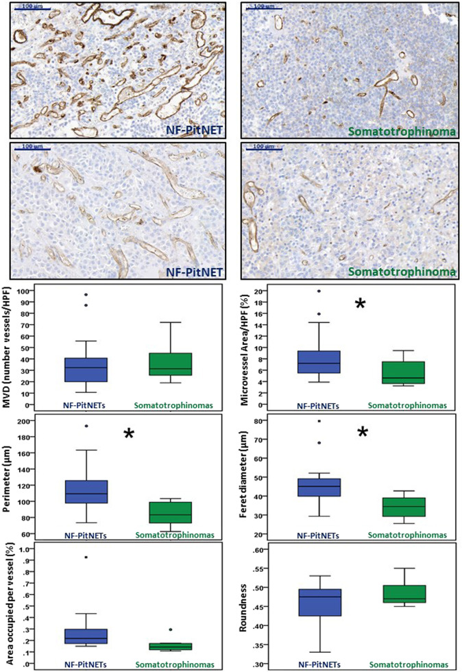
Fig. 2.
Angiogenesis in NF-PitNETs and somatotrophinomas. Microvessel density (MVD) and vasculature architecture parameters differences between human non-functioning pituitary neuroendocrine tumours (NF-PitNETs) and somatotrophinomas are shown. NF-PitNET (n = 16) and somatotrophinoma (n = 8) tissue sections were stained for CD31. CD31 positive vessels were counted in three different high-power fields (HPF) to obtain MVD (number of vessels/HPF). CD31-stained ×20 magnification fields were analysed with ImageJ and vessel contour was manually traced in order to obtain the vasculature architecture parameters: total microvessel area, area occupied per vessel, vessel perimeter, vessel Feret’s diameter and roundness (vessel roundness correspond to a value comprised between 0 and 1, with 1 = perfect circle). Representative images of vessels from two NF-PitNETs and two somatotrophinomas are shown (×20). Scale bar 100 µm. *p < 0.05, **p < 0.01, ***p < 0.001 (Mann–Whitney U test)

