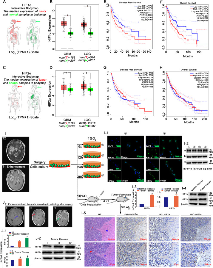
Fig. 1. Effects of HIF1α/HIF2α on the survival of patients with GBM.
a–d Both HIF1α and HIF2α were expressed at high levels in GBM; however, only HIF1α showed higher expression in tumours than in normal tissues. e–h Higher HIF1α expression led to shorter OS and DFS, and no significant differences were observed in OS and DFS between the higher and lower HIF2α expression groups. i GBM cells cultured in the presence of 1% O2 for 6, 12, 24 and 48 h exhibited increased HIF1α expression, but HIF2α levels were steadily maintained (I.1–I.2). The results revealed higher levels of HIF1α in tumour tissues, but a statistically significant difference in HIF2α levels was not observed between tumour and normal tissues (I.3–I.5). HypoxyprobeTM-1 detection verified the location of glioma in a hypoxic microenvironment (I.5). j HIF1α levels increased from WHO II to WHO IV grade tumours, but no significant difference in HIF2α levels was observed. All values are presented as the means ± SD. *P < 0.05 and #P > 0.05 were determined using Student’s t test or one-way analysis of variance, and the survival time was analysed using the log-rank test.

