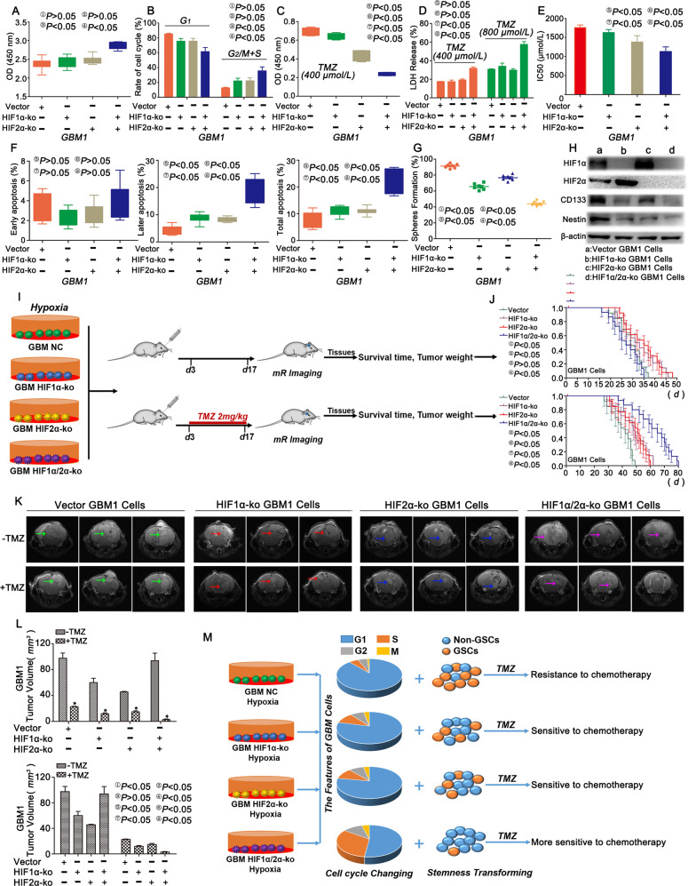
Fig. 3. Simultaneous HIF1α and HIF2α knockout increased proliferation and chemosensitization.
a No significant difference in proliferation was observed in GBM1 cells with single HIF1α or HIF2α knockout, but a higher cell proliferation rate was observed in cells with simultaneous HIF1α and HIF2α knockout in the absence of the TMZ treatment. b GBM1 cells with single HIF1α or HIF2α knockout presented no significant differences compared with the control, but a significant difference was observed after simultaneous HIF1α or HIF2α knockout, as this group presented the lowest percentage of cells in G1 phase compared with the other groups cultured in the presence of 1% O2. c HIF1α- or HIF2α-KO GBM1 cells exposed to TMZ (400 μM) for 72 h showed decreased proliferation, and the lowest proliferation rate was observed after simultaneous HIF1α and HIF2α knockout. d Higher levels of LDH release were observed in simultaneous HIF1α- and HIF2α-KO cells than in other cells. e The IC50 value decreased significantly after HIF1α or HIF2α knockout. f The percentages of late and total apoptotic cells increased after HIF1α or HIF2α knockout, and the highest percentage of apoptotic cells was observed in the group with simultaneous HIF1α and HIF2α knockout, but no significant difference was observed in the percentage of early apoptotic cells among groups. g A lower sphere formation rate was observed after HIF1α or HIF2α knockout. h CD133 and Nestin expression decreased after HIF1α or HIF2α knockout in cells. HIF1α expression increased after HIF2α knockout. In contrast, HIF2α expression increased after HIF1α knockout. i Schematic of the in vivo assay. j–l Analyses of the survival time and tumour volume in control and mice implanted with HIF1α/HIF2α-KO cells and treated with or without TMZ (2 mg/kg). m HIF1α or HIF2α knockout alone did not exert significant effects on proliferation and the cell cycle because of substitution effects, but inhibited stemness, leading to chemosensitization after TMZ treatment. However, if HIF1α and HIF2α were knocked out simultaneously, they inhibited cell cycle arrest, promoted proliferation, and decreased stemness, resulting in the chemosensitization of GBM cells. *P < 0.05 was determined using Student’s t test, and the specific P values are shown in Fig. S5E.

