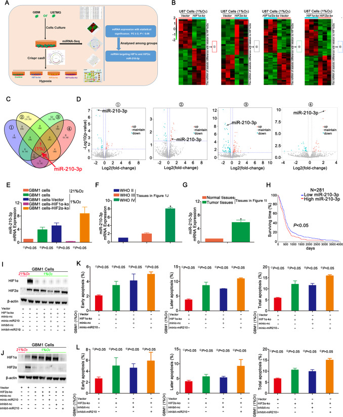
Fig. 4. miR-210-3p regulated HIF1α and HIF2α expression in hypoxic cells.
a Schematic of the mechanistic study. A miRNA-Seq analysis of HIF1α-KO, HIF2α-KO, simultaneous HIF1α- and HIF2α-KO and control cells was performed and revealed statistically significant differences in the expression of miRNAs targeting HIF1α or HIF2α in this process. b–d Heat maps showed statistically significant changes in the expression of miR-210-3p associated with HIF1α and HIF2α expression. The expression of miR-210-3p decreased in HIF1α-KO cells compared with control cells; however, its expression increased after HIF2α knockout. Compared with the simultaneous HIF1α and HIF2α knockout group, the control group exhibited increased miR-210-3p expression. Finally, for the HIF1α-KO and HIF2α-KO groups, higher miR-210-3p expression was observed in the cells of the HIF2α-KO group. e The expression of miR-210-3p was detected in GBM1 cells after HIF1α and HIF2α knockout using RT-qPCR. f Higher miR-210-3p expression was observed in WHO grade IV tumours compared with other tumour grades. g Higher miR-210-3p expression was observed in tumour tissues than in normal tissues. h TCGA database showed a lower survival time in the group with higher miR-210-3p expression. i, j Changes in HIF1α and HIF2α expression were detected in HIF1α- or HIF2α-KO GBM1 cells overexpressing or silencing for miR-210-3p and cultured in the presence of 1% O2. k, l Apoptosis was detected in HIF1α- or HIF2α-KO GBM1 cells overexpressing or silencing for miR-210-3p expression and cultured in the presence of 1% O2. *P < 0.05 was determined using Student’s t test, and the specific P values are shown in Fig. S6D.

