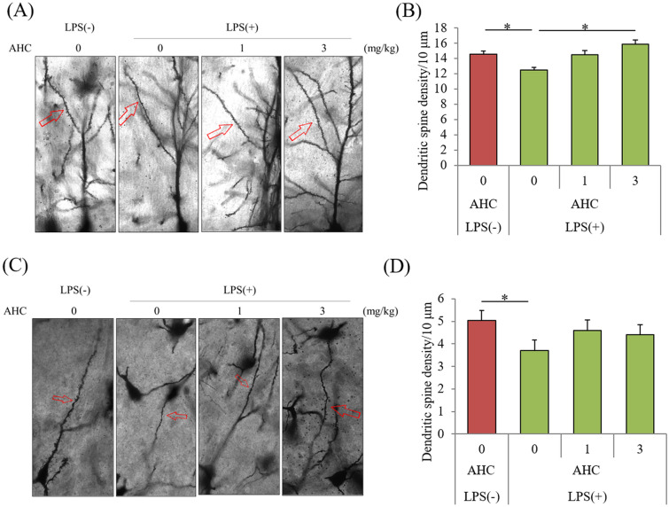
Figure 4.
Effects of the β-tricarbonyl moiety compound on LPS-induced changes in dendritic spine density. Crl:CD1 mice were orally administered with 0, 1, or 3 mg/kg of AHC for 3 days, and intracerebroventricularly injected with 10 μg of LPS. At 3 days after LPS treatment, the brains were subjected to Golgi staining. (A) and (C) Representative images of Golgi staining in CA1 of hippocampus and locus coeruleus (LC), respectively. (B) and (D) Number of dendritic spines per 10 μm in CA1 (B) and LC (D). Data are presented as mean ± standard error (7 mice per group). The p values shown were calculated by one-way analysis of variance followed by the Tukey–Kramer test. *p < 0.05. AHC, 2-acetyl-3-hydroxy-2-cyclopenten-1-one; LPS, lipopolysaccharide.

