Figure 2.
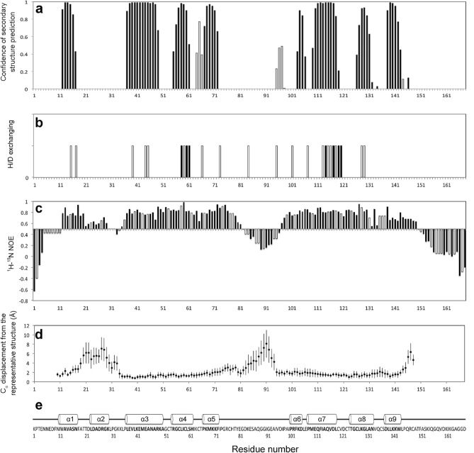
Residue-resolved structural and dynamics features of GLuc. (a) GLuc’s Secondary structure predicted by TALOS + : α-helix and β-sheet tendency are shown with solid and open bars, respectively. (b) H/D exchange experiments. Residues that retained resonance signal after incubation in D2O after 20 min and 18 h are marked with open bars and solid bars, respectively (1H–15N HSQC figures are shown in Supplemental Fig. 7). (c) 1H–15N heteronuclear NOE experiment data used to assess GLuc backbone flexibility: 1H–15N heteronuclear NOE are shown with solid bars. The NOE values of residues that were not identified were assumed using the average value of the preceding and following residues are shown with open bars. Flexible regions of GLuc were identified with the threshold value of 0.5. (d) Backbone Cα displacement from the representative structures is calculated using nineteen NMR-derived structures, and the error bars show standard deviations. (e) GLuc’s amino acid sequence and secondary structure as assigned in the representative structure.

