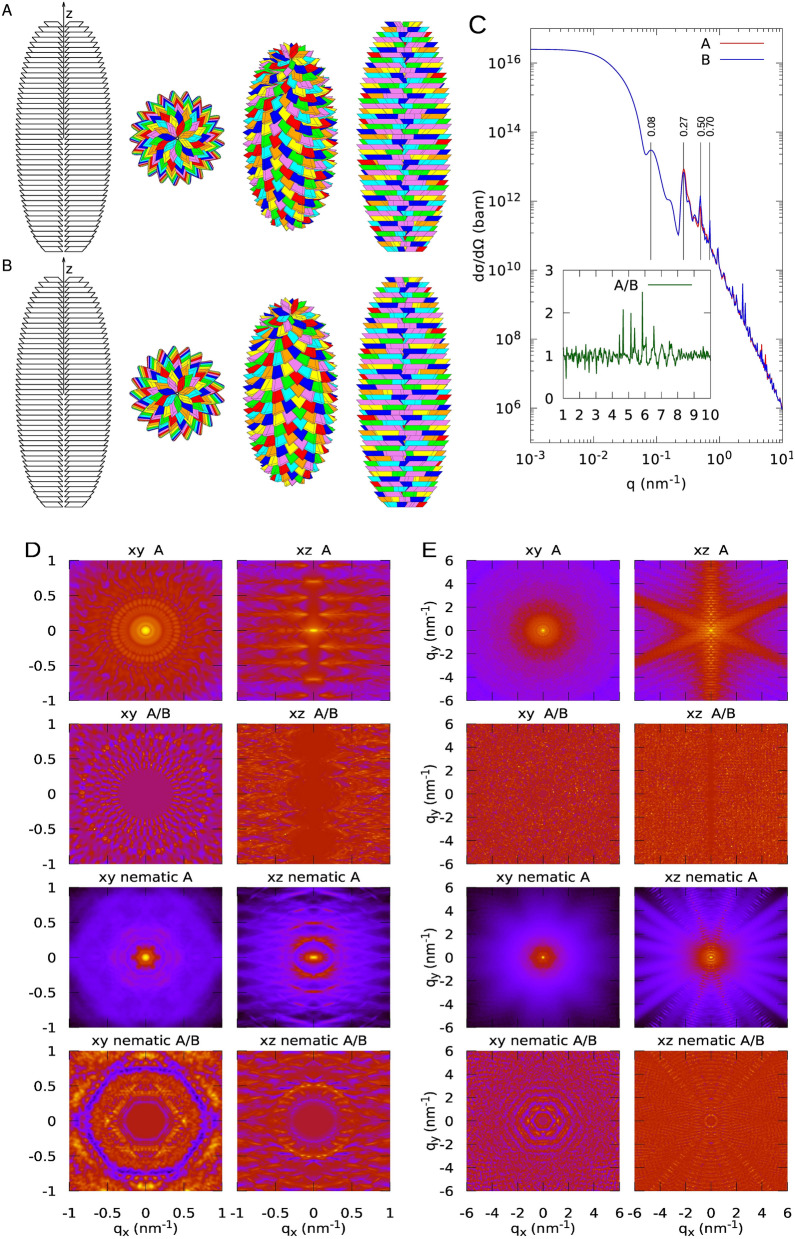
Figure 2.
Different views of a GSE according to the present model. Case #1. Geometrical parameters are: , nm, , , , , , nm, nm, nm, nm, . The number of layers in both bottom and top truncated cones is 23, and the volume fraction is . The overall molecular weight of the GSE is GDa. A. The first view on the left shows the inner cavity: platelets are not rotated by but only stacked for clarity. The other three pictures from left to right have been drawn by rotating the GSE along the axis by 0°, 45° and 90°, respectively. The platelets of each of the seven spirals are represented with the same colour: red, orange, yellow, green, cyan, blue and violet. B. GSEs obtained with the same parameters of panel A but with the specular images of the parallelepiped. C. SAXS and WAXS (highlighted in the inset) isotropic differential scattering cross-section of a GSE with direct and specular platelets, corresponding to panels A and B, respectively. D. Two-dimensional SAXS maps of the GSEs represented in panel A or B. The X-ray beam is assumed to be along the axis. The and views refer to the GSE with its long axis parallel or perpendicular to the X-ray or neutron beam. The first four maps refer to the GSE fixed in the space. The last four maps refer to GSEs arranged according to a nematic order, with order parameter , assuming that the director of the nematic phase is parallel or perpendicular to the X-ray or neutron beam ( nematic and nematic, respectively). E. WAXS maps corresponding to panel D.

