Figure 1.
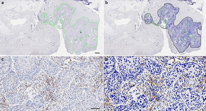
Immunohistochemical staining (IHC) and measurement of CD8+ tumor-infiltrating lymphocytes (TILs). (a) Low magnification image with an indication of tumor area by a solid green line. Areas of cystic space, non-specific stain, and dust were excluded from analysis (scale bar = 1 mm). (b) Converted image of 1A for the analysis scanned by Aperio (Leica Biosystems). (c) Representative image of high magnification. Many lymphocytes within the tumor area were stained with IHC staining for CD8 (scale bar = 50 μm). (d) Representative Aperio image of 1C. Lymphocytes are marked with blue (negative), yellow (1 +), orange (2 +), or brown (3 +) depending on staining intensity.

