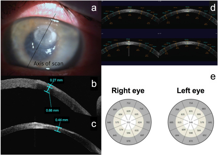
Figure 1.
Anterior segment optical coherence imaging protocol for bacterial keratitis (a–c) and controls (d, e). (a) Picture of bacterial keratitis, illustrating the anterior segment optical coherence tomography (AS-OCT) imaging protocol with the same axis high resolution scan carried out at presentation and after resolution of infection. (b) An AS-OCT scan at presentation, illustrating the measurement of corneal thickness (810 μm) and infiltrate thickness (270 μm) at presentation. (c) Measurement of final corneal thickness (440 μm) once the infection has resolved. (d) Four-quadrant AS-OCT scans of the control healthy cornea. The flap tool was used to identify a central 4 mm area, a mid-peripheral area defined by the 4 mm zone and an outer 8 mm zone, and a peripheral area extending from the 8 mm zone to the limbus. The corneal thickness was measured in the centre of each area on all four scans with calliper tools and the mean corneal thickness of all patients plotted. (e) The plotted corneal thickness maps of healthy control subjects.

