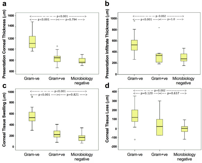
Figure 2.
Comparison of AS-OCT quantification parameters between Gram−ve, Gram+ve and microbiology negative bacterial keratitis. (a) There was a significant difference in presentation corneal thickness between Gram−ve and Gram+ve groups, and between Gram−ve and microbiology negative groups, but not between Gram+ve and microbiology negative groups. (b) There was a significant difference in presentation infiltrate thickness between Gram−ve and Gram+ve BK, and Gram−ve and microbiology negative BK, but not between Gram+ve and microbiology negative BK (p = 1.0). (c) There was a significant difference in corneal tissue swelling between Gram−ve and Gram+ve groups, and Gram−ve and microbiology negative groups, but not between Gram+ve and microbiology negative groups. (d) There was a borderline significant difference in corneal tissue loss between Gram−ve and Gram+ve BK (p = 0.12), a significant difference between Gram−ve and microbiology negative BK, and no significant difference between Gram+ve and microbiology negative BK.

