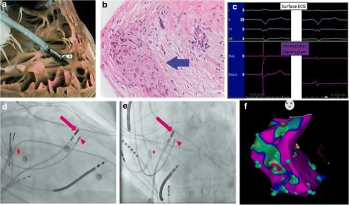
Fig. 3.
Endomyocardial biopsy of cardiac sarcoidosis. a Traditional endomyocardial biopsy with a bioptome. b Noncaseating granuloma suggesting a diagnosis of cardiac sarcoidosis. c The abnormal electrogram (right) is lower in voltages and fractionated. d, e RAO and LAO views of EGM-guided biopsy (red arrow, mapping catheter; red triangle, bioptome; star, intracardiac echocardiography). f Voltage map of patient with sarcoidosis created during biopsy with blue points representing abnormal bipolar signals. d, e Reproduced with permission from Liang et al. [37].

