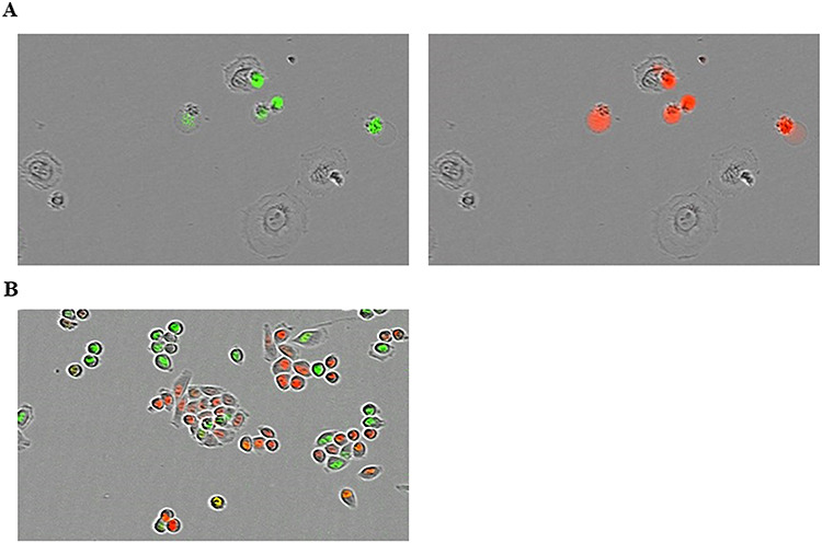
Fig. 1.

(A) Example of the fluorescence of apoptotic EMT6 cells (green) and cell death (red) irradiated at 2 Gy by X-rays without cisplatin. In this image, some cells fluorescing in red also fluoresce in green; in these cells, it is considered that apoptosis led to death of the cells. (B) Example of the fluorescence of the HeLa/Fucci2 cell cycle. Red and green cells are in the G1 and S/G2/M phases, respectively.
