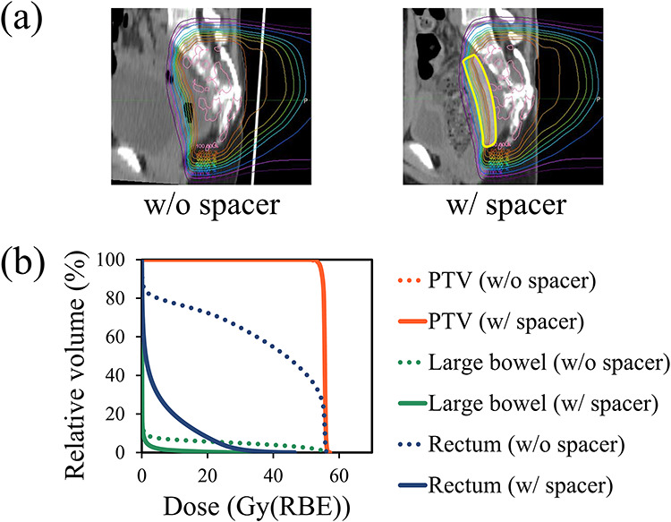
Fig. 2.

Plan comparison with (w) and without (wo) the PGA spacer in a pediatric patient. (a) Plans depicted on axial images at the same anatomical level with and without the PGA spacer. The thick yellow line indicates a contour of the PGA spacer. These dose distributions are normalized using the prescribed dose. (b) Comparison of DVHs for the PTV (red), large bowel (green) and rectum (blue) from the treatment plans with and without the spacer.
