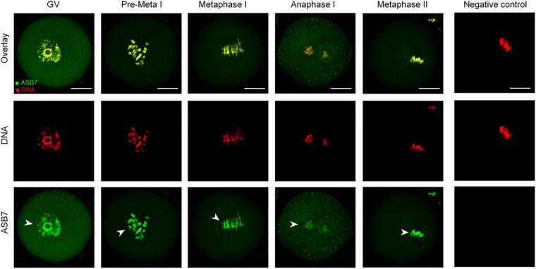
FIGURE 1.
ASB7 subcellular localization in mouse oocyte. Oocytes at GV, Pre-Metaphase I, Metaphase I, Anaphase I, and Metaphase II stages were immunolabeled with ASB7 antibody (green) and counterstained with propidium iodide (PI; red) for DNA. Negative control without primary antibody was also included. Representative images were acquired under confocal microscopy. Arrowheads show the accumulation of ASB7 signal. Scale bar: 30 μm.

