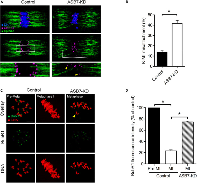
FIGURE 4.
Loss of ASB7 in oocytes impairs kinetochore–microtubule (K–MT) attachments. (A) Control and ASB7-KD metaphase oocytes were labeled with CREST for kinetochores (purple), α-tubulin antibody for microtubules (green), and Hoechst 33342 for chromosome (blue). Representative confocal images are shown; arrowheads show misattached kinetochores. Scale bar: 10 μm. (B) Quantitative analysis of K–MT attachments in control (n = 48) and ASB7-KD (n = 55) oocytes. Kinetochores in regions where fibers were not easily visualized were not included in the analysis. (C) Control and ASB7-KD oocytes were immunolabeled with anti-BubR1 antibody (green) and counterstained with PI to examine chromosomes (red). Representative confocal images of pre-metaphase I and metaphase I oocytes are shown. Arrowhead indicates the scattered chromosome in ASB7-KD oocytes. Scale bar: 10 μm. (D) BubR1 fluorescence intensity in control (n = 46) and ASB7-KD (n = 42) oocytes was quantified. Data are expressed as mean percentage ± SEM of three independent experiments. *p < 0.05 vs controls.

