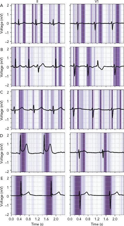
Figure 4: Important Regions for the Deep Neural Network to Predict Whether an ECG is Normal, Abnormal or Acute.

ECG leads II and V1 with a superimposed guided Grad-CAM visualisation showing regions important for the deep neural network to predict whether an ECG is normal, abnormal or acute. A and B: Normal ECGs with focus on the P wave, QRS-complex, and T wave, while correctly ignoring a premature ventricular complex. C: Abnormal ECG with a long QT interval and a focus on the beginning and end of the QT-segment. D and E: Acute ECGs with an inferior ST-segment elevation MI (D) and a focus on the ST-segment and with a junctional escape rhythm (E) and a focus on the pre-QRS-segment, where the P wave is missing. Source: van de Leur et al. 2020.[19] Reproduced from the American Heart Association, Inc., by Wiley Blackwell under a Creative Commons (CC BY-NC-ND 4.0) licence.
