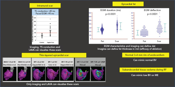
Figure 4: Problems with Field of View.

Top to bottom, left to right: intramural scar can be missed by voltage mapping. Pacing techniques can help to detect such scars. Epicardial fat can mimic low BV, but has less duration and deflections than fibrotic scar. Subepicardial scar from myocarditis can be missed by conventional UV and BV mapping. Manual reannotation can help to demask these thin layers. Small rims of normal endocardial voltage above the scar can hide the substrate. Tissue oedema during RF can make ME EGM disappear due to their limited field of view. BV = bipolar voltage; EGM = electrogram; LAVA = local abnormal voltage activity; ME = mini- or micro-electrodes; RF = radiofrequency; TS = transseptal; UV = unipolar voltage.
