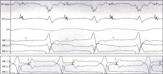
Figure 5: Example of Difference in Field of View.

Patient with ischaemic cardiomyopathy and ongoing stable ventricular tachycardia (VT). Mapping with Intella MiFi catheter shows sharp, large, fragmented mid-diastolic signal, suggesting a VT isthmus. This signal that was clear on radiofrequency distal was not observed on the mini-electrodes (ME), which are embedded in the tip electrode. Movement of few millimetres to improve contact suddenly showed the same local electrogram on the ME, where VT is terminated successfully. This states the difference in field of view of conventional ablation electrodes and the importance of contact force. Source: P Maury. Reproduced with permission from P Maury.
