
Figure 1.
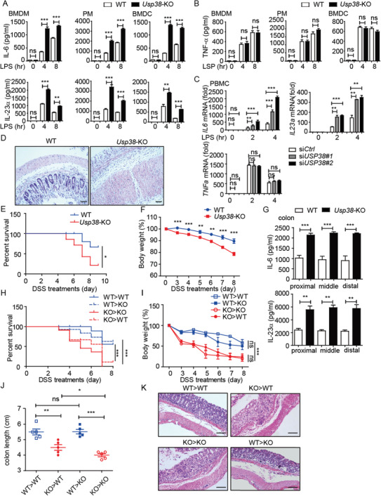
Loss of USP38 enhances inflammatory responses in vitro and in vivo. A) Protein levels of IL‐6 and IL‐23α in BMDMs, BMDCs, and PMs from wild‐type (WT) and Usp38‐knockout (KO) mice during LPS stimulation for the indicated time points. B) Protein level of TNF‐α in BMDMs, BMDCs, and PMs from WT and Usp38‐KO mice during LPS stimulation for the indicated time points. C) Expression level of IL6, IL23a, and TNFa mRNA in PBMCs transfected with USP38‐specific siRNAs (siUSP38#1, siUSP38#2) or control (siCtrl) siRNA under LPS treatment for the indicated time points. D–F) WT and Usp38‐KO mice were challenged with 2.5% dextran sulfate sodium (DSS) for 7 d and sacrificed at day 8. KO, n = 6; WT, n = 6. Histopathology of the distal colon (D), survival (E), and weight changes (F) in WT and Usp38‐KO mice. G) Protein levels of IL‐6 and IL‐23α in colon tissue from (D). H–K) Survival, weight changes, colon length, and histopathology of the distal colon in the indicated bone marrow chimera mice (n = 8–12/group). Data in (A)–(C) are presented as the means ± SEM of at least three biological experiments. Data in (E)–(G) are presented as the means ± SD of the experiment with six mice per group. Data in (H)–(J) are presented as the means ± SD of the experiment with 8–12 mice per group. Data in (D)–(K) are representative of three independent experiments. *p < 0.05, **p < 0.01, ***p < 0.001, ns, no significant difference, versus the wild type or control group with the same treatment (Student's t‐test in (A)–(C), (G), (F), (I), (J) and Mantel–Cox test and Gehan–Breslow–Wilcoxon test in (E) and (H)).