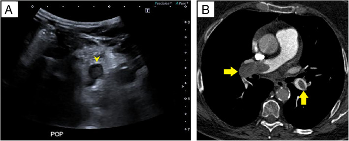
Figure 1.

A, Thrombosed popliteal vein. Transverse ultrasound scan shows an echogenic clot in the left popliteal vein (arrowhead). B, Bilateral PE. Computed tomographic pulmonary angiography shows bilateral filling defects in the right pulmonary artery and the left lower lobar artery (arrows).
