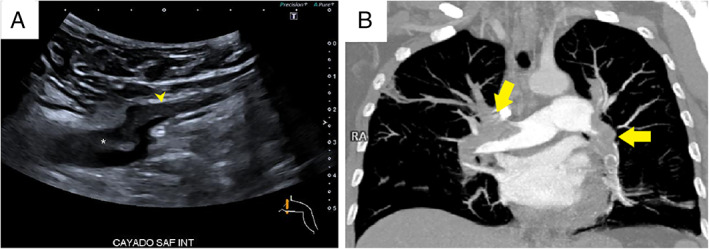
Figure 2.

A, Thrombus in the common femoral vein (CFV). Transverse ultrasound scan shows a partial filling defect in the saphenous vein (arrowhead) and the common femoral vein (asterisk) just above the saphenous junction. B, Bilateral pulmonary embolism. Coronal maximum‐intensity projection CT pulmonary angiography shows bilateral filling defects in the two main pulmonary arteries and their lobar branches (arrows).
