Figure 2.

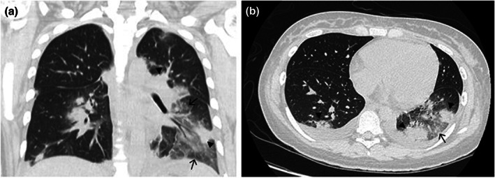
Chest computed tomographic scans 36 h after surgery. Ground‐glass opacity and a crazy‐paving appearance (arrows) with consolidation (arrow heads) in the bilateral lungs. (a) Frontal plane. (b) Transverse plane.

Chest computed tomographic scans 36 h after surgery. Ground‐glass opacity and a crazy‐paving appearance (arrows) with consolidation (arrow heads) in the bilateral lungs. (a) Frontal plane. (b) Transverse plane.