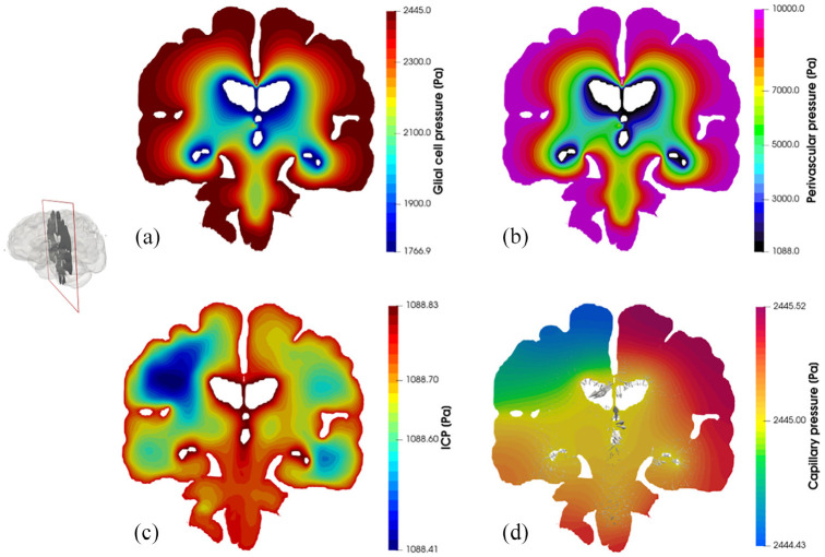
Figure 4.
Coronal slices of the 6-MPET solution fields allied to the parenchymal volume of the 72-year-old cognitively healthy female subject undergoing a period of high activity. Both the pore pressure of the glial cell compartment (a) and the perivascular/glymphatic system (b) is symmetric in morphology, reflecting the nature of the boundary conditions imposed for these compartments whilst also considering the intercompartmental influences of the remaining compartments. The latter influences are more prevalent in the compartments describing the (c) intracranial pressure (CSF/ECF compartment) and (d) capillary pressure, since the former superimposes a highly distinctive permeability tensor map which can capture the underlying parenchymal microstructure (see Guo et al. and Vardakis et al.47,48 for more details). The capillary compartment and its accompanying pore pressure reflect the asymmetric nature associated with the arterial flow variability being applied on the partitioned parenchymal surface, as it is in direct communication to the high transmantle pressure gradient present in that compartment. The superimposed velocity vectors associated to the paravascular/glymphatic system filtration velocity assist in attenuating the level of detail associated with this rich dataset.

