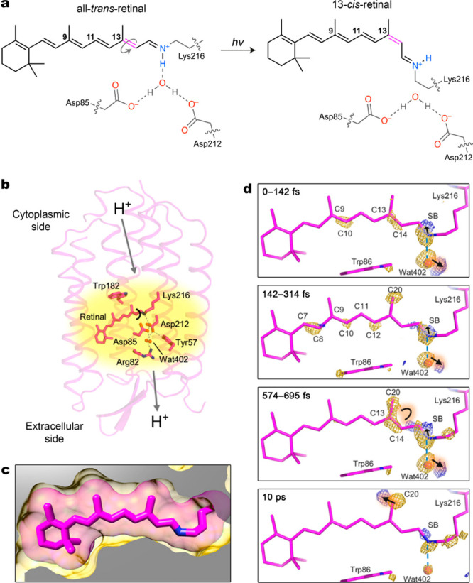
Figure 1.

(a) Light-induced regioselective isomerization of retinal within the binding pocket of bacteriorhodopsin. (b) Structure of bacteriorhodopsin with retinal buried inside its hydrophobic cavity. The arrows indicate the direction of light-induced proton transfer. (c) Binding of retinal (pink sticks with van der Waals radii shown as a transparent halo) encased within the hydrophobic cavity of bacteriorhodopsin (yellow). (d) Structural dynamics of retinal and its immediate surroundings captured by a femtosecond X-ray laser. The transition from trans-retinal to cis-retinal is mapped onto a dark-state model based on the difference Fourier electron density (Fobslight – Fobs) contoured at 4σ (yellow, negative; blue, positive). Adapted with permission from ref (5). Copyright 2018 American Association for the Advancement of Science.
