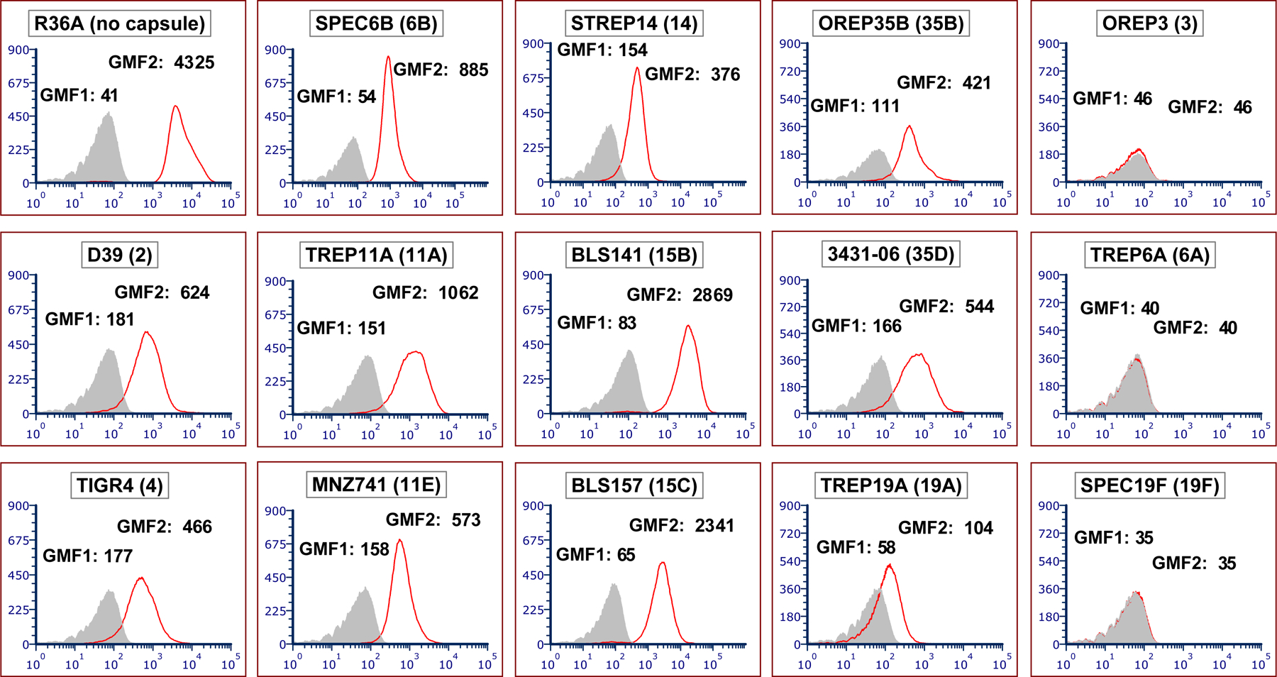
FIGURE 1.

DBA binds to a majority of pneumococcal strains by flow cytometry. Flow cytometric histograms showing binding of DBA to different target strains. Strain names are indicated at the top of each panel, with the associated serotype in parentheses. The y-axis indicates the number of events per channel and the x-axis indicates fluorescence intensity. The grey-filled areas were obtained with bacteria co-incubated with fluorescein-conjugated streptavidin (secondary antibody) only, and this background signal is represented as geometric mean fluorescence 1 (GMF1). The red line represents DBA binding for each strain, with bacteria co-incubated with biotin-conjugated DBA (primary antibody) and fluorescein-conjugated streptavidin, and this signal intensity is represented as GMF2
