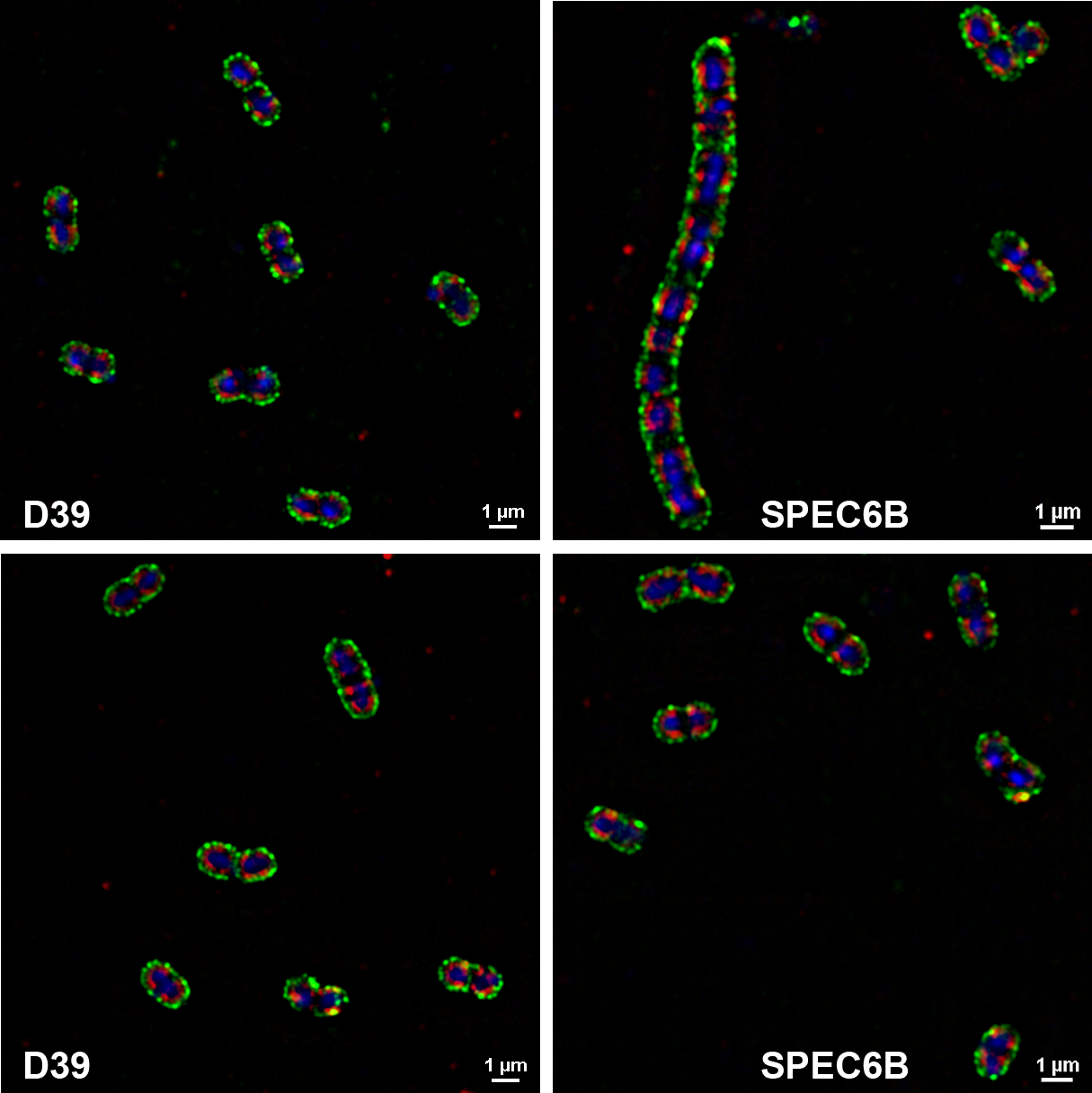
FIGURE 2.

Visualization of capsule and DBA staining with super-resolution structured illumination microscopy. D39 and SPEC6B were stained with DBA (red) and anti-capsule antibodies (green; Hyp2M2 for D39 and Hyp6BM1 for SPEC6B). DNA was stained with DRAQ5 (blue)
