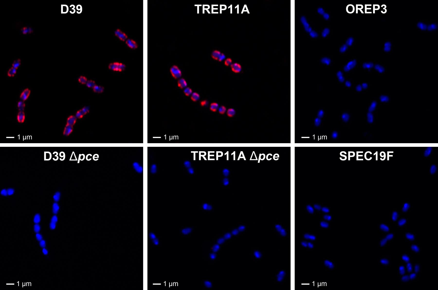
FIGURE 7.

Visualization of HPA binding to various pneumococcus strains using confocal fluorescence microscopy. Six representative strains, including D39 (pce allele A), TREP11A (pce allele B), their corresponding pce knock-out mutants, and two strains with no Pce activity (OREP3 and SPEC19F) are shown for HPA staining. Red represents HPA staining and blue represents DNA counter-staining using DRAQ5
