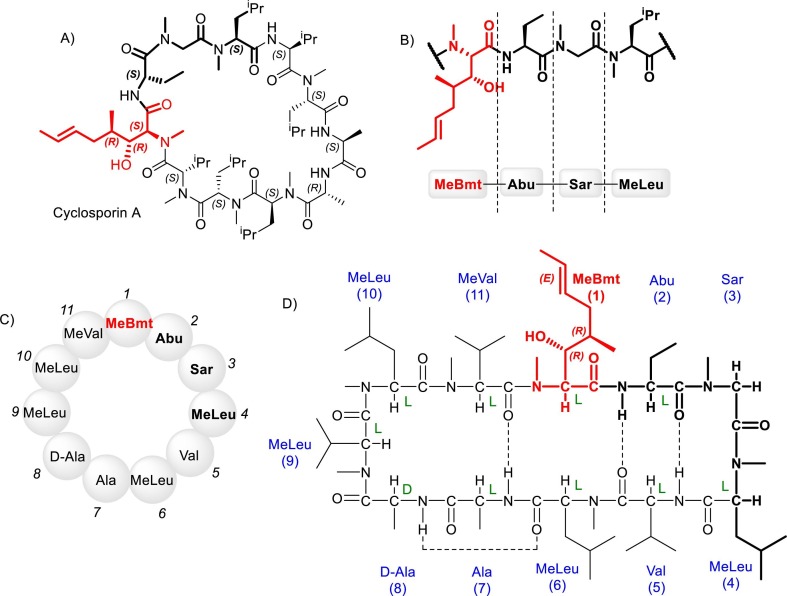
Fig. 19.
Different representations for the structure of cycloporin A. (a) Angular chemical representation. (b) Amino acid residues sequence associated with the drug's pharmacodynamic interaction. (c) Peptide primary structure representation. d) Linear representation highlighting the intramolecular hydrogen bonds. MeBmt = (4R)-4-((E)-2-butenyl)-4-methyl-L-threonine residue; Abu = α-aminobutyric acid residue; Sar = sarcosine residue; MeLeu = N-methylleucine residue; MeVal = N-methylvaline residue; Ala = alanine residue; Val = valine residue; - - - - = Hydrogen bonds.

