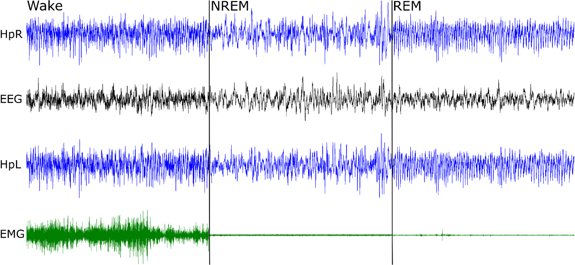
Figure 6. Recording from the headplate.

Right hippocampus (HpR), electroencephalogram (EEG), left hippocampus (HpL) and electromyogram from the neck muscle (EMG) were recorded. The total duration of the panel is 30 seconds, with 10 second examples of wake, non-REM sleep and REM sleep. Note sequential fall of EMG amplitude and prominent theta activity (here ~9 Hz), best seen in hippocampal electrodes during REM sleep.
