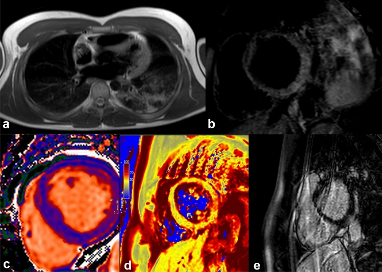
Fig. 2.
A 27-year-old patient, without any significant past medical history, was admitted to our hospital with fever and chest pain. The onset of symptomatology dated back about 1 week. His initial investigation showed elevated troponin levels at laboratory tests. Electrocardiography displayed ST-segment elevation. Viral myocarditis of unknown aetiology was initially suspected, but SARS-Co-V-2 as a cause was ruled out later at serology. Echocardiography was normal. A chest X-ray showed pulmonary consolidation at the left lower lobe. Cardiac magnetic resonance imaging confirmed the myocarditis (panel a). T2 STIR (panel b) showed an increased signal in mid-basal inferior and inferior-lateral segments. The analysis of T1 mapping (panel c) showed an increase in signal at the same segments (average values of 1100 ms, with reference values of 1030 ± 30 ms). T2 mapping values (panel d) showed an increased signal in mid-basal inferolateral segment (65 ms. Reference values: 52 ± 3 ms), thus indicating the presence of edema. In the sequences acquired later after contrast, an area of sub-epicardial LGE in mid-basal inferior and infero-lateral segments was observed with a concomitant involvement of the adjacent pericardium (panel e). Images processed with Circle CVI 42

