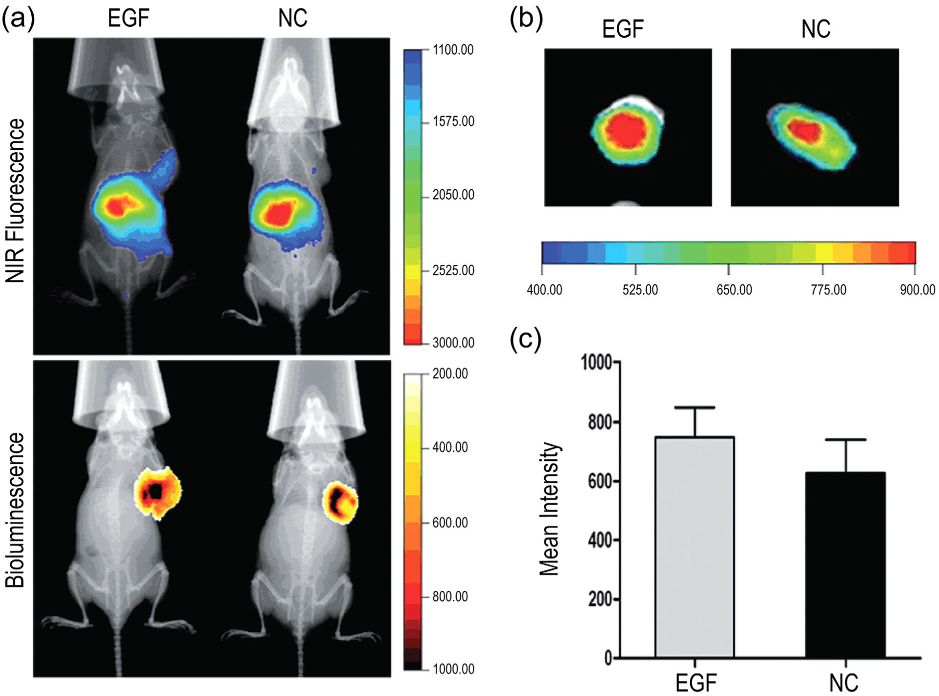
FIGURE 10.

In vivo and ex vivo imaging of tumor-bearing mice treated with nonconjugated (NC) or EGF-conjugated dendriplexes (EGF). (a) Upper panel: localization of EGFR-targeted and untargeted dendrimers via near infrared (NIR) fluorescence at 2 hr posttreatment. Lower panel: luminescence imaging of the localization of breast cancer tumor cells labeled with luciferase (MDA-MB-231-Luc), implanted subcutaneously, and following administration of 150 mg/kg D-luciferin. (b) Overlay of X-ray and ex vivo NIR fluorescence images of tumors treated with NC or EGF-conjugated dendriplexes. (c) Mean ± SD ex vivo fluorescence intensity of tumors 2 hr posttreatment with NC or EGF-conjugated dendriplexes (J. Li et al., 2016)
