
Figure 14 |. trans-Tango visualizes neurons postsynaptic of JO-A and JO-B neurons.
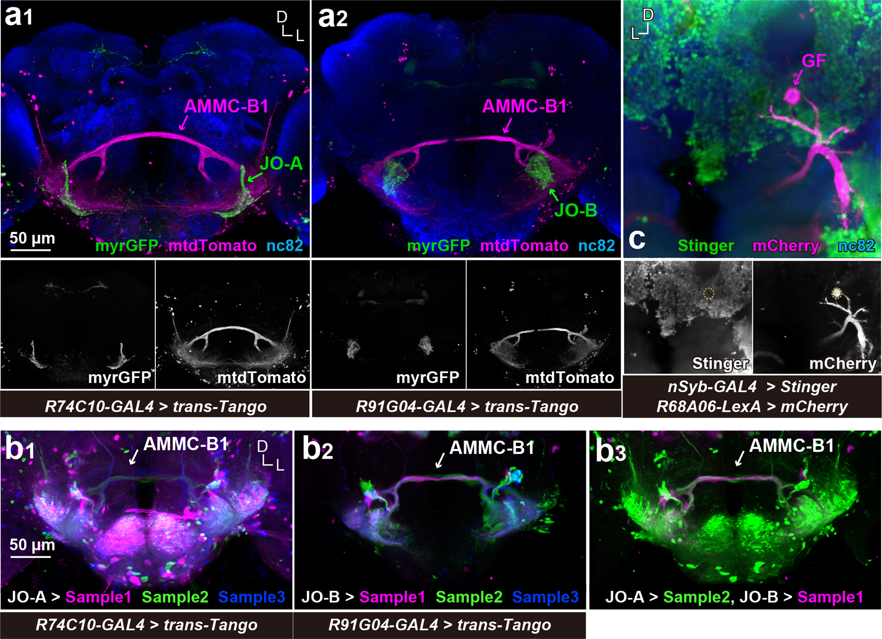
(a) AMMC-B1 neurons are monosynaptic downstream of JO-A and JO-B neurons. R74C10-GAL4 (a1) and R91G04-GAL4 (a2) drivers label JO-A and JO-B neurons, respectively (green). trans-Tango using these GAL4 drivers was used to visualize AMMC-B1 neurons (magenta). Nc82 antibodies visualize neuropil in the brain. Stacked images of optical sections are shown, in which anterior and posterior sections of the brain are not included for clarity. (b) trans-Tango signals after image registrations. (b1 and b2) Three trans-Tango brain samples for JO-A (R74C10-GAL4) (b1) and JO-B neurons (R91G04-GAL4) (b2) are shown. Three samples registered to a single brain space are overlaid in different colors. (b3) Overlay of JO-A and JO-B trans-Tango brains. The trans-Tango signals of JO-A and JO-B are the same as those in the left (JO-A > Sample 2) and middle panels (JO-B > Sample 1), respectively. (c) GF neuron is devoid of nSyb-GAL4 signals. GF neuron is labeled using mCherry-HA marker (magenta). Nuclei of nSyb-GAL4 positive cells are labeled with Stinger, a nuclear-localized GFP marker (green). The cell body of the GF neuron is outlined in the bottom panel. D, dorsal; L, lateral.