
Figure 4 |. EM tracing of AMMC-B1 and GF neurons.
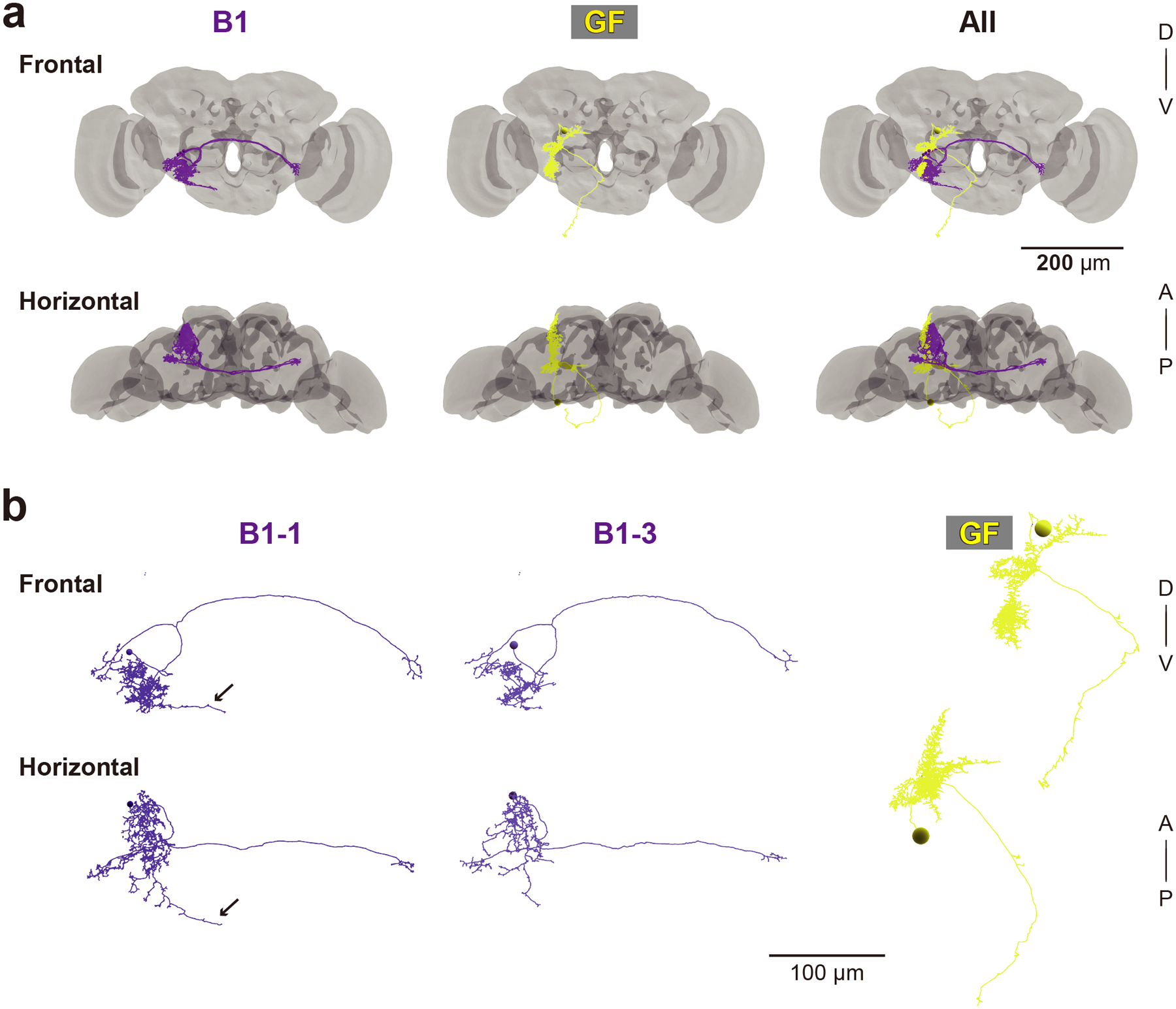
(a) 3D reconstructed images of five AMMC-B1 neurons (B1, dark violet) and a GF neuron (GF, yellow). Gray backgrounds represent the neuropil of a fly brain (Zheng et al., 2018). (b) Reconstruction of AMMC-B1 and GF neurons. Arrows indicate the AMMC-B1 branches innervating the GNG (left). Each AMMC-B1 neuron is named serially (B1–1 to B1–5), in which only typical examples (AMMC-B1–1 and B1–3) are shown (left and middle). A, anterior; D, dorsal; P, posterior; V, ventral.