Figure 1.

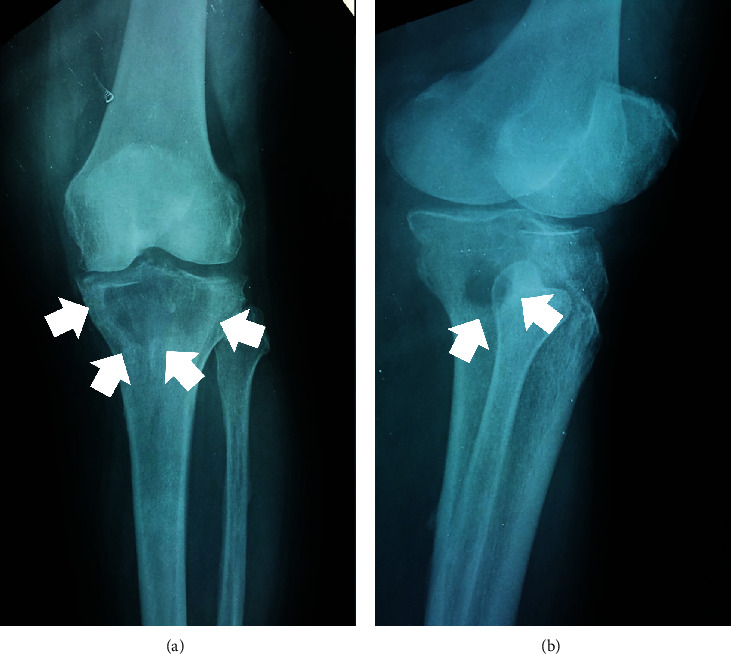
Preoperative radiograph ((a) anteroposterior, (b) lateral) of the left knee, depicting the existence of a large lytic lesion (white arrows) at the proximal tibia, extending into the popliteal cavity.

Preoperative radiograph ((a) anteroposterior, (b) lateral) of the left knee, depicting the existence of a large lytic lesion (white arrows) at the proximal tibia, extending into the popliteal cavity.