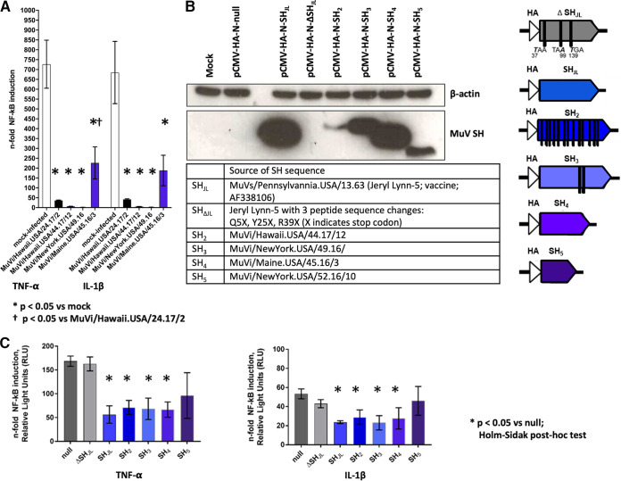
FIG 2.
(A) A549 cells were transfected (X-tremeGENE HP DNA transfection reagent, Roche) with pCMV-β-Gal and p(PRDII)5tkΔ(−39)lucter, which carries a luciferase gene expressed under the control of an NF-κB-inducible promoter. Transfected A549 cells were inoculated at 24 h posttransfection with MuVs at an MOI of 0.5 and, 16 h later, were stimulated with either TNF-α or IL-1β or were not stimulated. Cell lysates were harvested 4 h poststimulation for quantification of luminescence and beta-galactosidase activity using commercially available assay systems (OneGlo Luciferase Assay System and Beta-Galactosidase Enzyme Assay System, respectively; Promega). Luminescence values from each sample were normalized based on beta-galactosidase activity and were expressed in relative light units (RLU). The fold change of NF-κB induction was estimated by calculating the ratio between RLU from stimulated versus unstimulated controls for each condition. The SH amino acid sequence of MuVi/Hawaii.USA/24.17/2 is identical to the reference strain MuVi/Sheffield/GBR/01.05 [G]; this condition was included as a positive control for SH-mediated abrogation of NF-κB signaling. Statistical analysis was performed by one-way ANOVA followed by post hoc Holm-Sidak correction for multiple comparisons in GraphPad Prism v8. (B) Noncanonical SH sequences were cloned for ectopic expression through high-fidelity assembly (NEBuilder HiFi Assembly Mix; New England BioLabs) into expression vector pCMV-HA-N. Plasmid constructs were verified by Sanger sequencing (BigDye v3.1, ABI). A549 cells were transfected with the indicated plasmids (X-tremeGENE HP DNA transfection reagent, Sigma-Aldrich) and lysates were harvested at 24 h posttransfection. Expression of N-terminally tagged SH proteins was confirmed by immunoblot following SDS-PAGE of lysates (20 μg total protein) in denaturing conditions (12% Bis-Tris protein gel in MES buffer, Invitrogen) and transfer to nitrocellulose using the iBlot system (Invitrogen). Membranes were probed for HA (H3663, Sigma-Aldrich) and endogenous β-actin (A3854, Sigma-Aldrich), which served as a loading control. Sources and characteristics of the cloned SH sequences are summarized in text and in graphic form, respectively. (C) A549 cells were cotransfected with reporter plasmids and either null vector or SH expression vectors, as described in panel A. Cells were stimulated with either TNF-α or IL-1β at 24 h posttransfection and then harvested for detection of luminescence and beta-galactosidase activity.

