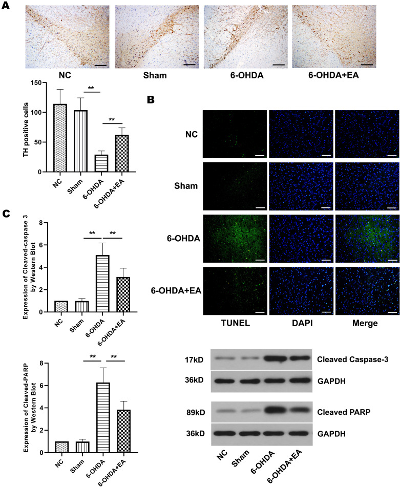
Fig. 3.
Effect of EA on neuronal apoptosis in the substantia nigra of 6-OHDA-induced rats. A. TH positive cells in the substantia nigra of rats from NC group, Sham group, 6-OHDA group, and 6-OHDA+EA group were stained after 6-OHDA lesioning. Scale bar=200 µm. B. Photomicrographs showed the TUNEL positive cells (green) in the substantia nigra of rats from each group. DAPI (2-4-Amidinophenyl-6-indolecarbamidine dihydrochloride) was used to stain nuclei (blue) for light microscopy. Scale bar=100 µm. C. Western blot showed the expression of cleaved caspase-3 and cleaved PARP in the substantia nigra of rats from each group. Data are expressed as mean ± SD. **P<0.01.

