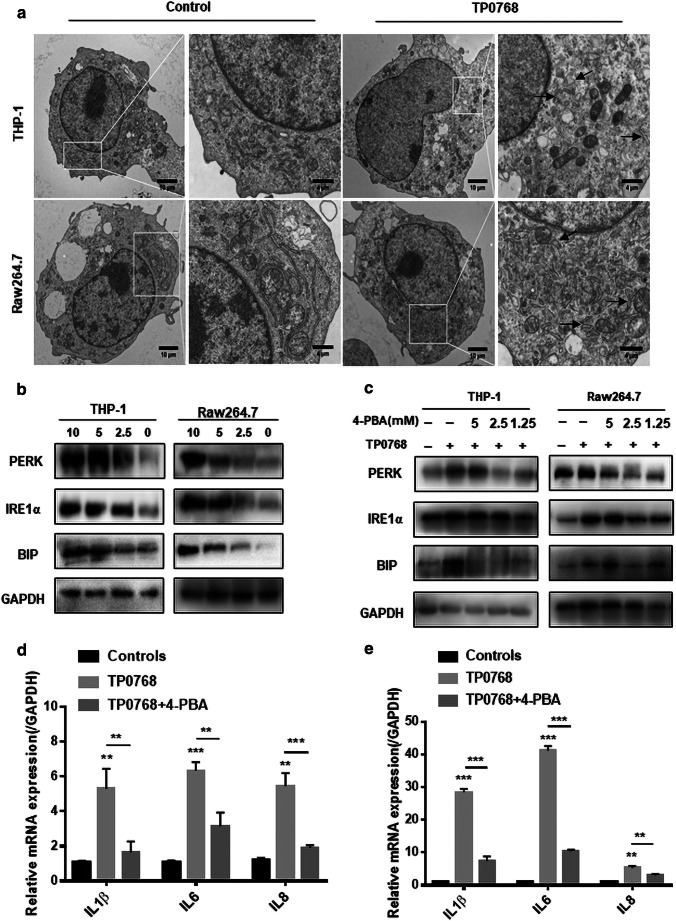
Fig. 2.
Tp0768 mediated the expression of IL-1β, IL-6, and IL-8 through ER stress. a Electron microscope image of the ultrastructure of THP-1–differentiated macrophages and Raw264.7 cells treated with 5 μg/mL Tp0768. The high-magnification image highlights the swollen endoplasmic reticulum. b After treatment of cells with different concentrations of Tp0768 for 24 h, ER stress-related proteins, PERK, IRE1α, and Bip, were detected using western blotting. c Cells pre-incubated with 4-PBA (5, 2.5, and 1.25 mM) for 1 h were treated with Tp0768 for 24 h, and the protein levels of PERK, Bip, and IRE1 were evaluated using western blotting. d, e After pretreatment with 4-PBA (2.5 mM) and co-treatment with Tp0768 for 24 h, the mRNA expression of IL-1β, IL-6, and IL-8 were evaluated using RT-qPCR. Values are expressed as fold changes relative to GAPDH-normalized mRNA levels. All data are presented as mean ± SD of at least three independent experiments. *p < 0.05, **p < 0.01, and ***p < 0.001 indicate a significant difference from the control group

