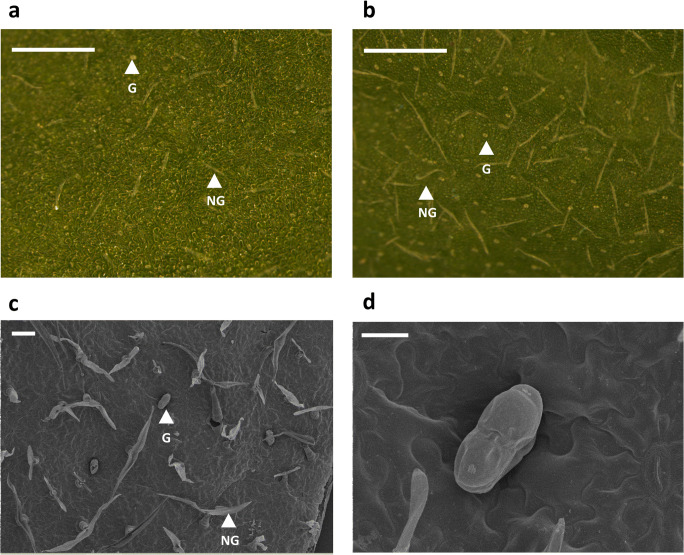
Fig. 1.
Representative micrographs of non-glandular and glandular trichomes on chrysanthemum leaves. Light micrographs of the adaxial leaf surface of a chrysanthemum cultivar displaying (a) low and (b) high trichome densities. Scanning electron microscopic images of the adaxial leaf surface of chrysanthemum leaves (c and d). Bean-shape glandular trichomes are shown in (d). White arrows indicate the position of non-glandular (NG) and glandular (G) trichomes. The white bars represent 100 μm in (a), (b) and (c), and 30 μm in (d)

