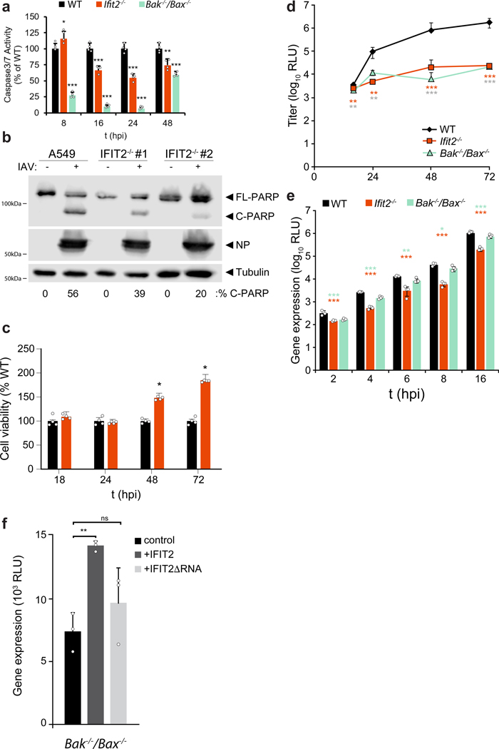
Extended Data Fig. 3. Human IFIT2 and murine Ifit2 promote virus-induced apoptosis.
a, Caspase 3/7 activity was measured in wildtype, Ifit2−/−, and Bak/Bax−/− MEFs infected with WSN at an MOI of 0.01 for the indicated times. n = 4 independent infections ± standard deviation. b, Expression of cleaved (C-PARP) versus full-length (FL-PARP) PARP-1 in IAV infected A549 and IFIT2 knockout cell lines. Protein bands were quantified and the percent C-PARP of total PARP signal normalized to a tubulin loading control is indicated below. c, Viability of Ifit2−/− cells during infection with WSN assessed by CellTiter Glo Assay at the indicated time points. n = 4 independent infections ± standard deviation. d, Multicycle replication of WSN reporter virus in in WT, Ifit2−/−, and Bak/Bax−/− MEFs. n = 3 independent infections ± standard deviation. e, Single-cycle WSN reporter virus gene expression at early time points post-infection in WT, Ifit2−/−, and Bak/Bax−/− MEFs. n = 4 independent infections ± standard deviation. f, WSN reporter virus gene expression in Bak/Bax−/− cells ectopically expressing an empty vector, hIFIT2, or hIFIT2-ΔRNA. n = 3 independent infections ± standard deviation. *p < 0.05, **p < 0.01; *** p < 0.001; Two-tailed Student’s T-test for pairwise comparisons (c) or one-way ANOVA with post hoc Tukey’s HSD for multiple comparisons to WT cells (a, d, e and f). All data are plotted as mean ± sd.

