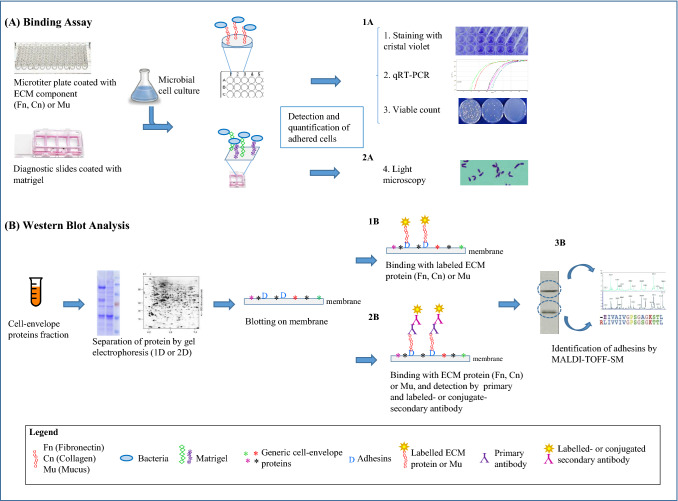
Fig. 1.
Graphic representation of the most common in vitro model systems described to study bacteria/host interaction. a Detection of bacterial adhesion to mucus (Mu) or ECM components, e.g. fibronectin (Fn) and collagen (Cn). Binding assay can be performed on microtiter plate, coated with one ECM component or mucus (upper), or on diagnostic slides coated with matrigel (lower), which contains mostly Cn and laminin. Microbial cell culture of the strain under study is added in each well and, after washing, adhered cells can be detected and quantified by different methods: 1a—staining with crystal violet [61], qRT-PCR [77] or viable count [49], when microtiter plate is used; 2a—by light microscopy, when diagnostic slides are used [73]. b Identification of proteins involved in the bacteria/host interaction. Extracted surface proteins are separated by mono-dimensional (1D) or two-dimensional (2D) gel-electrophoresis and western blotted by using labeled ECM or mucus components [9] (1b), or specific polyclonal antibodies and labelled or conjugate secondary antibody [77] (2b). Identification of putative adhesins may be obtained by MALDI-TOF Mass Spectrometry (3b)

