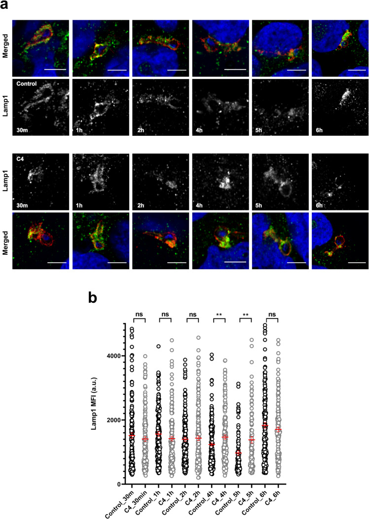
Fig. 5. C4 increases lysosomal marker protein Lamp1 colocalization with the liver-stage PVM.
Huh7 cells infected with P. berghei parasite were treated with 1 µM C4 and fixed at different time point post-compound addition. DMSO (0.0001%) was used as the negative control. a Representative confocal images of P.berghei infected Huh7 cells treated with C4/DMSO. Cells were stained with anti-UIS4 (red), anti- Lamp1 (green) and Hoechst (blue). Scale bars = 5 µm. b Quantification of Lamp1 intensity around the PVM. The graph represents Lamp1 mean fluorescence intensity (MFI) around the PVM marked by UIS4. The data represents means ± SEM (n = 2 independent experiments). N ≥ 100 parasites. P values were calculated using unpaired two-tailed t-test. ns: non-significant, **P < 0.01.

