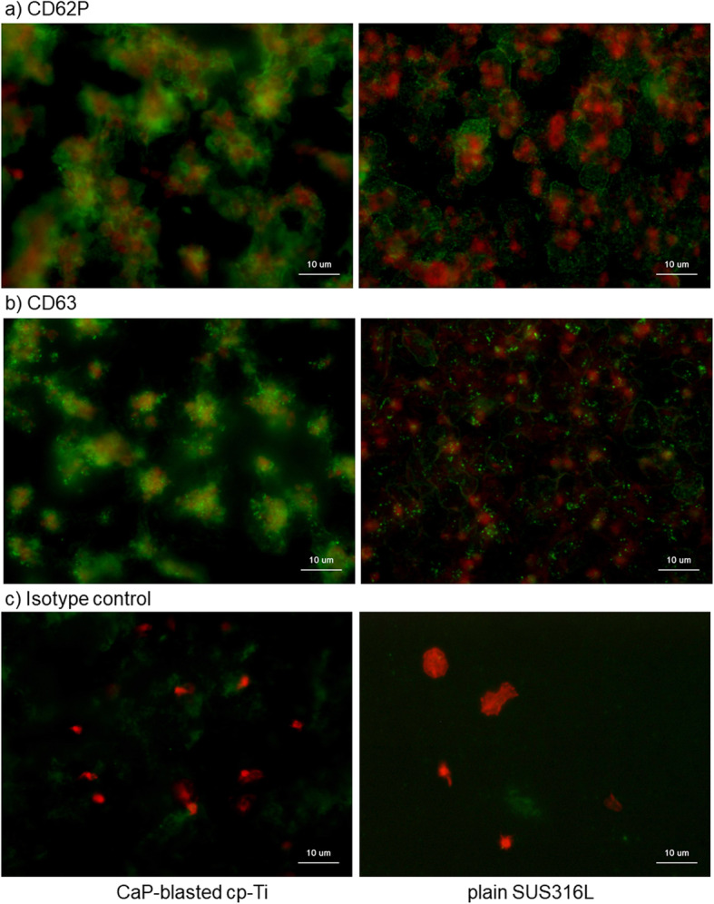
Fig. 9.

Immunocytochemical detection of activated platelets expressing a CD62P or b CD63. CaP-blasted cp-Ti and plain SUS316L plates were incubated with PRP for 60 min, washed, fixed, and subjected to immunocytochemical visualization. Green represents proteins reactive for a CD62P or b CD63, while red represents polymerized actin. c To show non-specific detection in extra-platelet spaces, PRP containing relatively low platelet counts was added on the plates and examined
