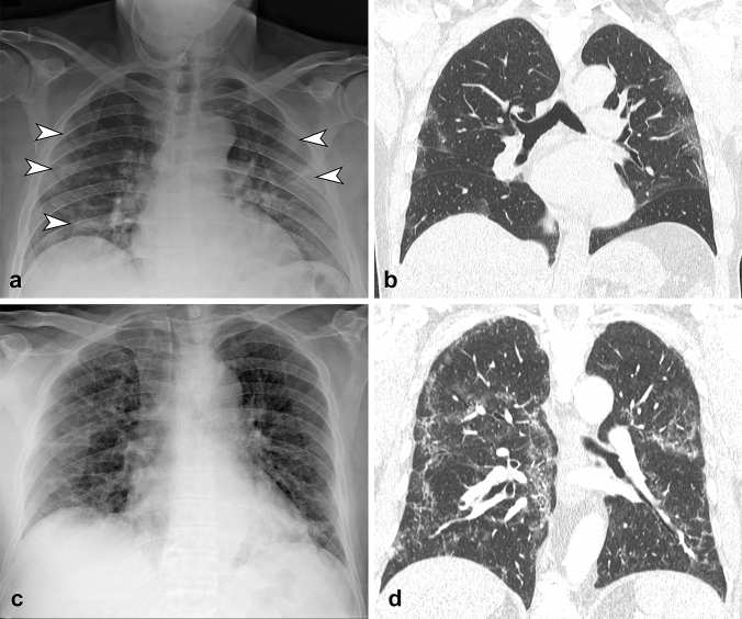
Fig. 1.
a CXR in antero-posterior view shows bilateral ground glass opacities (arrowheads) with peripheral distribution, involving middle-lower zone of the lungs. b Coronal reformatted CT image confirmed the CXR findings. c CXR in antero-posterior view shows bilateral reticular pattern with diffuse distribution on both axial and longitudinal plane. d Coronal reformatted CT image confirms the presence of diffuse interstitial involvement with reticular pattern

