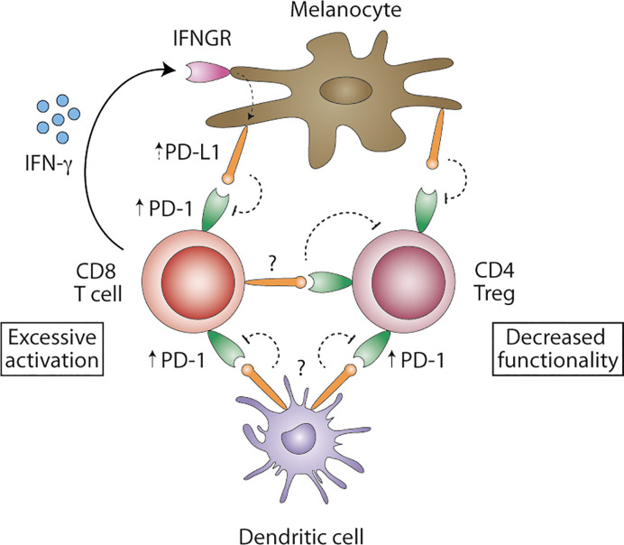
Figure 1.
Proposed model of PD-1/PD-L1 signaling in vitiligo. Blood regulatory CD4+ and cytotoxic CD8+ T cells show increased levels of PD-1 protein. IFN-γ-producing CD8+ T cell are abundantly present in affected skin and may induce PD-L1 expression on melanocytes, albeit possibly ineffective in inhibiting autoreactive T cells. Consequently, melanocyte-reactive CD8+ T cells remain activated. PD-L1 expression on DCs remains unstudied in vitiligo and might be important in effectively inhibiting autoreactive CD8+ T cells. PD-L1+ DC might inhibit not only T cell priming in the tumor-draining lymph nodes, but also T cell effector function. PD-L1 expression in situ remains unclear but might be expressed by melanocytes, DC or melanocyte-specific T cells, turning PD-1+ regulatory CD4+ T cells to functional exhaustion. Overcoming excessive activation of autoreactive CD8+ T cells and decreased functionality of regulatory CD4+ T cells in vitiligo might be achieved by interfering with PD-1/PD-L1 signaling.

