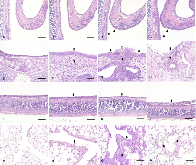
Fig. 3.
Histopathology of rats exposed to PHMG·HCl. In the nasal cavity, (A, B) No abnormal lesion was observed in the control (A) and 1 mg/m3 PHMG·HCl (B) groups. (C) Degeneration of respiratory epithelium (arrow) and transitional epithelium (arrowhead) in the 5 mg/m3 PHMG·HCl group. (D) Squamous metaplasia of respiratory epithelium (arrow) and transitional epithelium (arrowhead) in the 25 mg/m3 PHMG·HCl group. In the larynx, (E) No abnormal lesion was observed in the control group. (F, G) Squamous metaplasia of the epithelium (arrow), inflammation of the lamina propria (arrowhead) in 1 (F) and 5 mg/m3 (G) PHMG·HCl groups and ulcer of the epithelium (white arrow) in 5 mg/m3 (G) PHMG·HCl group. (H) Ulcer of the epithelium (arrow) and inflammation of the lamina propria (arrowhead) in the 25 mg/m3 PHMG·HCl groups. In the trachea, (I) No abnormal lesion was observed in the control group. (J, K) Degeneration of the epithelium (arrow) in 1(J) and 5 mg/m3 (K) PHMG·HCl groups. (L) Necrosis of the epithelium (arrow) in the 25 mg/m3 PHMG·HCl group. In the lung, (M) No abnormal lesion was observed in the control group. (N) Detachment of the bronchiolar epithelium (arrow) in the 1 mg/m3 PHMG·HCl group. (O) Squamous metaplasia of bronchiolar epithelium (arrow) in the 5 mg/m3 PHMG·HCl group. (P) Alveolar fibrosis (arrow) in the 25 mg/m3 PHMG·HCl group. Scale bars=100 μm, Magnification: ×200, H&E staining.

