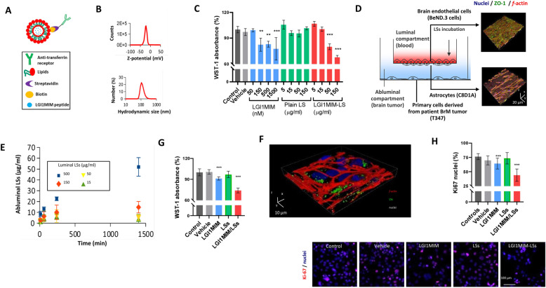
Fig. 5.
The anti-proliferative effect of LGI1MIM on metastatic tumour and the anti-cancer effect and potential of LGI1MIM-loaded liposomes (LGI1MIM/LSs) for crossing the blood–brain barrier (BBB). a Schematic of LGI1MIM/LSs functionalised with the antibodies against transferrin receptor. b Z-potential and hydrodynamic size (diameter) of the LGI1MIM/LSs. c WST-1 cell viability assay reveals the anti-proliferative effects of both LGI1MIM and LGI1MIM/LSs (single administrations; 72 h of treatment) on primary cells derived from brain metastatic tumour (T347). Mean ± SD *p < 0.05; **p < 0.01; ***p < 0.001, ANOVA HSD post hoc test is used. d Schematic of the multicellular 2D model of the BBB (left); 3D confocal imaging of the endothelial layer (top right) and of the astrocytes (bottom right); nuclei in blue, zonula occludens-1 (ZO-1) in green and f-actin in red. e BBB crossing of DiO-stained LGI1MIM/LSs incubated in the luminal compartment at increasing concentrations (15, 50, 150 and 500 μg/ml). LGI1MIM/LSs were detected in the abluminal compartment. f 3D confocal laser scanning microscopy imaging of T347 cells after treatment with 500 μg/ml LGI1MIM-LSs (single administration in the luminal compartment; 72 h of incubation). g WST-1 cell viability assay on T347 cells in response to a single administration in the luminal compartment of vehicle, 5 μM LSs, 500 μg/ml LGI1MIM, or 500 μg/ml LGI1MIM-LSs (72 h of incubation) *p < 0.05; **p < 0.01; ***p < 0.001, ANOVA HSD post hoc test is used. h Ki-67 expression on T347 cells in response to the treatments reported in (k). *p < 0.05; **p < 0.01; ***p < 0.001, ANOVA HSD post hoc test is used

