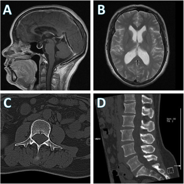
Fig. 1.
Brain magnetic resonance imaging (MRI) and lumbar computed tomography (CT) performed during the patient’s first admission. a Brain MRI demonstrated mild enlargement of the supratentorial ventricle, b the abnormal sign of vacuolar sella in the right optic-radiation of lateral thalamus, bilateral medial temporal lobes, and the insular lobes. c, d No abnormalities were found in lumbar CT at first; however, a retrospective review of the spinal CT scan (d) showed the evidence of enlarged neural foramina and mild vertebral scalloping which suggested a long-standing intradural tumor such as schwannoma

