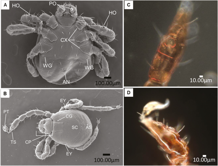
Fig. 2.
Representative Scanning Electron Microscope images of larvae of Rhipicephalus microplus identifying several different sensory organs taken by Brenda Leal and Alejandra Fuentes using a Hitachi TM4000 Scanning Electron Microscope. (A) ventral view; (B) dorsal view: papal organs( PO), Haller’s organ (HO), wax glands (WG), coxa (CX), anus (AN), scutum (SC), cervical groove (CG), alloscutum (AS), capitulum (CP), eyes (EY), pretarsus (PT), and tarsus (TS); and (C and D) Close up of the Haller’s organ taken by Dr. Donald B. Thomas using a Keyence VHX-7000 digital microscope.

