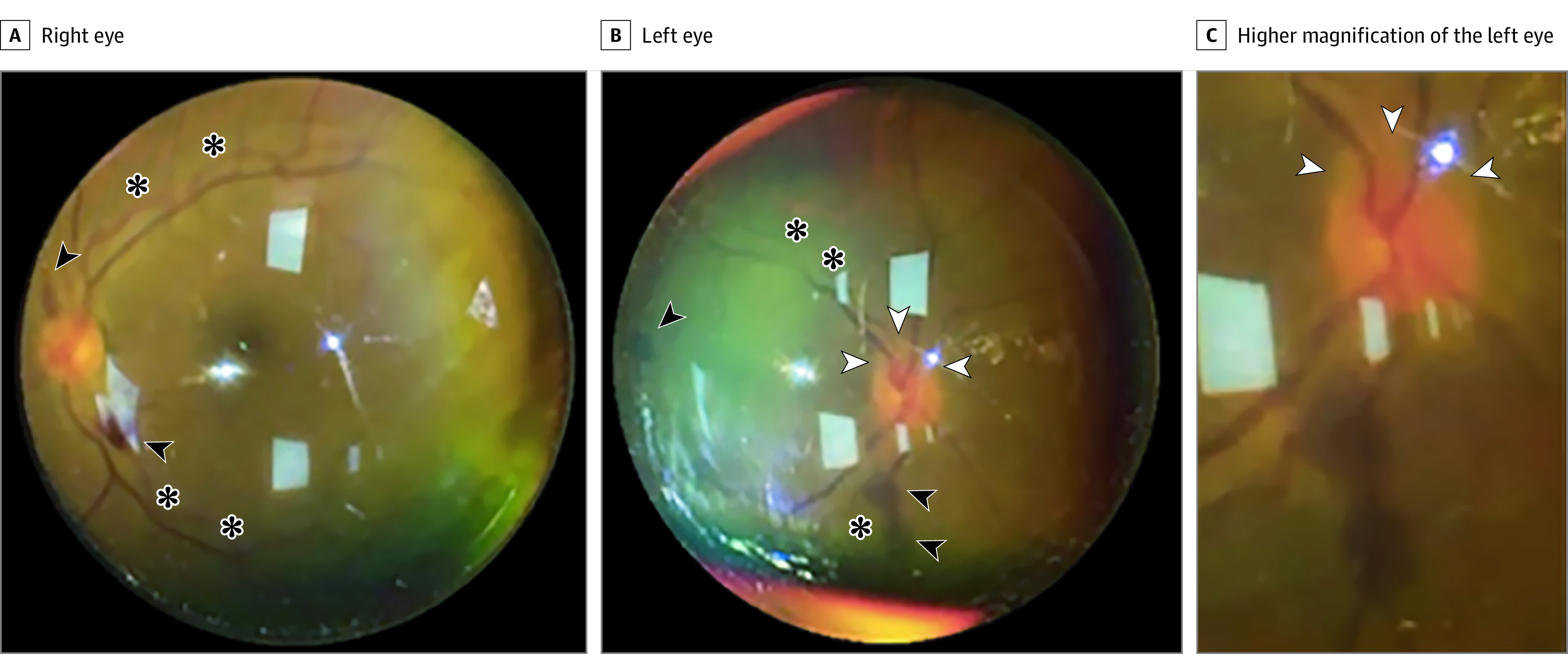
Figure 1. Case 1: A Man in His Early 50s After 9 Sessions of 18-Hour Prone-Position Ventilation.

Fundus photograph taken with an iPhone 6S and 20-diopter lens at the intensive care unit bedside. The right eye (A) optic disc had sharp margins with nerve fiber layer hemorrhages (arrowheads) superiorly and inferiorly near the optic disc. There was tortuosity (asterisks) of the retinal veins. The left eye (B) optic disc had mild inferior margin elevation (white arrowheads), with a few intraretinal and subretinal hemorrhages (black arrowheads) in the midperiphery. There was tortuosity (asterisks) of the retinal veins. C, Higher magnification of the left optic disc showed mild inferior margin elevation (arrowheads).
