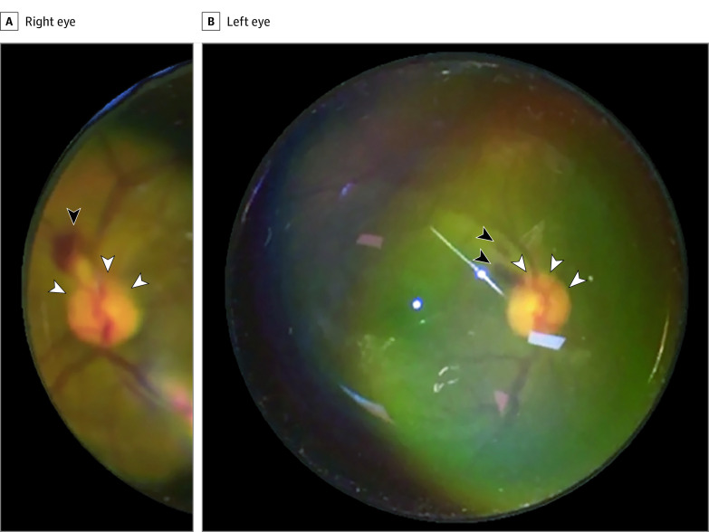
Figure 2. Case 2: A Man in His Mid-40s After 4 Sessions of 18-Hour Prone-Position Ventilation.
Fundus photograph taken with an iPhone 6S and 20-diopter lens at the intensive care unit bedside. The right eye (A) and left eye (B) had inferior optic disc elevation (white arrowheads) with associated flame-shaped hemorrhages (black arrowheads).

